Combination of rays
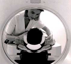
 x-rays have been put to enormous use in medical imaging since Roentgen first obtained a picture of the bones in his hand, almost a hundred years ago. But in the last few years, several new tools like magnetic resonance imaging ( mri ), computer aided tomography ( cat ) and positron emission tomography ( pet ) have been added to the tools with which we can see the human body.
But now for the first time there are reports of combining two complementary techniques. Researchers at the University of California in Los Angeles, us , have succeeded in getting the first ever simultaneous pet and mri images of a sample. The problem has been the incompatibility of the very powerful magnetic fields used by the mri scanners and the sensitive electronics needed for pet images. The pet scanner typically uses a detector array made up of crystals of a compound of bismuth, germanium and oxygen ( bgo ). This detector absorbs the gamma rays given out by the tracer and gives out a burst of photons, which are then captured by a bank of photomultiplier tubes. These amplify the signal which is then converted into an electrical signal and processed by a computer. The trouble with bgo detectors is that they have a moderately good resolution and so the group used a new material called lso (lutetium, silicon and oxygen). This provided a much better resolution since it emits many more photons than bgo .
The next problem was to isolate the electronics of the pet scanner from the mri magnets. This was done by connecting thin optical fibers to the back of the detector and piping the photons to a safe distance away from the magnets. The group led by Simon Cherry has tested the apparatus with images of a plastic cylinder with holes filled with a radioactive substance. Though many more improvements in the size of the apparatus and its resolution will have to be achieved before it becomes useful to image humans, the successful test of the prototype is a promising achievement
These imaging techniques have created a revolution in our understanding and the treatment of the human body. Magnetic resonance imaging uses very powerful magnetic fields to probe the vibrational properties of the hydrogen atom in the water molecules present in all the tissues of the body. The image is a very high-resolution map, which has all the anatomical details but carries no direct indication of the functional properties of tissues.
pet on the other hand uses a radioactive tracer which is injected into the body. The tracer is absorbed by active cells of the body and it subsequently gives out gamma rays. The rays are detected by an array of crystal detectors and the information is fed into a computer after being processed by intermediate electronic circuits. The pet images are very good functional maps because they can distinguish between living and dead tissues, by the differential rates of absorption of the radioactive tracer. ( Science , Vol 272, Vol 5267).
Related Content
- An overview of major shark and ray catchers, traders, and species
- Wildfires and geochemical change in a subalpine forest over the past six millennia
- In a first, Narendra Modi govt gives 600 MW stranded power projects second wind
- Attenuation of urban agricultural production potential and crop water footprint due to shading from buildings and trees
- Ooty model of bio-remediation to save urban waterbodies
- UP to add 1,100MW toits power kitty today
