
New eye on organ formation
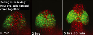 a new study on how eyes develop in a growing embryo overturns the textbook model of the process leading to organ formation. It found that cells that form an organ usually migrate to the place where the organ will be located early on in development rather than the entire tissue in its proximity being responsible for its creation.
Conducted by Jochen Wittbrodt and his lab at the European Molecular Biology Laboratory in Heidelberg, Germany, the study used advanced microscope techniques to track individual cells in the transparent embryos of a small fish called Medaka.
In 2001, Felix Loosli from Wittbrodt's laboratory discovered a protein called Rx3 that is required for eye formation. Only cells that will become the eye produce this molecule. For the study, Martina Rembold, also from Wittbrodt's group, labelled these cells with a fluorescent marker and tracked them using advanced software developed by Richard Adams at the University of Cambridge. The team found that single cells migrate actively and one-by-one from the centre of the brain to form eyes. The study appeared in the August 25 issue of Science (Vol 313, No 5790).
Related Content
- Report by the Kerala State Pollution Control Board regarding Periyar river pollution, 19/11/2024
- Improving access to the Green Climate Fund: how the fund can better support Developing Country institutions
- Affidavit by the Government of NCT of Delhi on steps taken to control Yamuna pollution and rejuvenate the river, 25/01/2021
- Order of the National Green Tribunal regarding coastal pollution on Marina beach, Chennai, Tamil Nadu, 08/09/2020
- Future heavy-duty emission standards: An opportunity for international harmonization
- Order of the National Green Tribunal regarding reconstitution of the Committee constituted by MoEF&CC for SLHEP, 31/07/2019